Scientists have developed contact lenses that can capture and detect exosomes, which could be cancer biomarkers.
The Terasaki Institute for Biomedical Innovation (TIBI) has developed a contact lens that can capture and detect exosomes, nanometre-sized vesicles found in bodily secretions, such as tears, which have the potential to act as a diagnostic cancer biomarker.
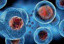
Exosomes are formed within most cells and secreted into bodily fluids such as urine and tears. They were believed to collect the unwanted materials from the original cells; however, exosomes can actually transport different biomolecules between cells. Exosomes possess proteins which increase in response to cancer, viral infections, or injury.
Can exosomes detect cancer biomarkers?
The capabilities of exosomes have sparked an ongoing interest in their abilities to act as a potential cancer biomarker, aiding in diagnosis and treatment prediction. Further research delving into this has impeded due to the challenging nature of isolating exosomes in sufficient quantity and purity.
Current methods to isolate exosomes are tedious and time-consuming as they involve ultracentrifuge and density gradients, lasting around 10 hours to complete. There are further difficulties in the detection of isolated exosomes, and commonly used methods require expensive and space-consuming equipment.
Developing smart contact lenses
The scientists developed the contact lenses to capture exosomes found in tears as a potential way to detect cancer biomarkers. They utilised their expertise in contact lens biosensor design and fabrication to eliminate the requirement of tricky isolation methods by creating their antibody-conjugated signalling microchamber contact lens (ACSM-CL). The technology captures exosomes from tears which is the cleaner and more optimum method to discover cancer biomarkers.
The team used alternative approaches when optimising the ACSM-CL; they used direct laser cutting and engraving rather than the conventional cast moulding for structural retention of both the chambers and the lens.
Additionally, the scientists incorporated a method that chemically modifies the microchamber surfaces to activate them for antibody binding. This method is an alternative to standard approaches, in which metallic or nanocarbon materials must be used in expensive clean-room settings.
The team optimised procedures for binding a captured antibody to the ACSM-CL microchambers and a different (positive control) detection antibody onto gold nanoparticles that are visualised spectroscopically. Both these antibodies are specific for two surface markers found on all exosomes.
During the initial validation experiment, the ACSM-CL was also tested against exosomes secreted into supernatants from 10 different tissue and cancer cell lines. The ability to capture and detect exosomes was validated by the spectroscopic shifts observed in all the test samples in comparison with the negative controls. Similar results were obtained when the ACSM-CL was tested against 10 different tear samples collected from volunteers.
In final experiments, exosomes in supernatants collected from three different cell lines with multiple types of surface marker expressions were tested against the ACSM-CL, along with various combinations of the marker-specific detection antibodies. The patterns of detection and non-detection of exosomes from the three different cell lines were as expected, validating the ACSM-CL’s ability to accurately capture and detect exosomes with different surface markers, including cancer biomarkers.
“Exosomes are a rich source of markers and biomolecules which can be targeted for several biomedical applications,” said Ali Khademhosseini, PhD, TIBI’s Director and CEO. “The methodology that our team has developed greatly facilitates our ability to tap into this source.”


