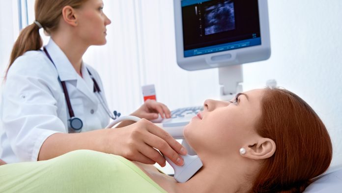
The University of Sheffield has developed a new ultrasound method that could help diagnose abnormal tissue, scarring and cancer.
A new ultrasound method that can measure the level of tension in human tissue for the first time, which is an indicator of diseases, has been developed by the University of Sheffield.
The breakthrough discovery, made by Dr Artur Gower from the University’s Department of Mechanical Engineering, together with researchers from Harvard, Tsinghua University, and the University of Galway, could be used to build new ultrasound machines that can better diagnose abnormal tissue, scarring, and cancer.
The research paper can be found in the Science Advances journal.
What are ultrasounds?
Ultrasounds use sound waves to create images of organs inside the human body. However, the current imagery produced is not typically enough to diagnose abnormal tissues.
The team set out to improve diagnosis by developing a way to measure forces such as tension by using an ultrasound machine. Tension is generated in all living tissue, so measuring it can indicate whether tissue is functioning properly or if it is affected by disease.
Harnessing a technique from a rail project
The researchers used a technique from a rail project at the University of Sheffield, which uses sound waves to measure tension along railway lines. The technique, used both for rail and medical ultrasound, relies on a simple principle of the ‘greater the tension, the faster sound waves propagate’. Using this principle, the researchers developed a method that sends two sound waves in different directions. The tension is then related to the speed of the waves by using mathematical theories developed by researchers.
Previous ultrasound methods have failed to show the difference between stiff tissue and tissue under tension. Therefore, this new technique is the first to measure the tension of soft tissue without knowing anything about it.
Dr Artur Gower, Lecturer in Dynamics at the University of Sheffield, said: “When you go to the hospital, a doctor might use an ultrasound device to create an image of an organ, such as your liver, or another part of your body, such as the gut, to help them explore what the cause of a problem might be. One of the limitations of ultrasounds used in healthcare now is that the image alone is not enough to diagnose whether any of your tissues are abnormal.
“What we have done in our research is developing a new way of using ultrasound to measure the level of tension in the tissue. This level of detail can tell us whether tissues are abnormal or if they are affected by scarring or disease. This technique is the first time that ultrasound can be used to measure forces inside the tissue, and it could now be used to build new ultrasound machines capable of diagnosing abnormal tissue and disease earlier.”


