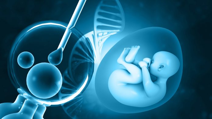
A team of scientists based in the UK have identified an exciting new method in which human embryos interact with their mother-to-be.
The team, comprised of researchers at The University of Manchester and Manchester University NHS Foundation Trust, conducted in vitro research that revealed a new way human embryos communicate with their mother-to-be after just six days of its development. In addition, the study illuminated that the embryos could detect foreign cells in their environment, including pathogens.
The research is published in the journal Human Reproduction today.
Novel capabilities of human embryos
The researchers found that when foreign cells are detected by the embryos’ special proteins called Toll Like Receptors (TLRs), they emit a type of protein called a cytokine. This cytokine – called Interleukin-8 (IL8) – typically plays a role in the body’s immune system by recruiting neutrophils to a site of inflammation. Despite this knowledge, its role in the relationship between human embryos and their mother was unknown until now.
The team examined the behaviour throughout the fifth and sixth days of the embryos’ development, a period where it is free-living in the female body, subsequently implanting in the lining of the uterus and beginning pregnancy. TLRs are understood to be found in the fallopian tube and vagina, which is known as the maternal tract. TLRs two and four are also known to be expressed by human sperm. However, there has not been similar investigations of human embryos.
Daniel Brison, the lead author of the study and an Honorary Professor at The University of Manchester and Scientific Director at the Department of Reproductive Medicine at Saint Mary’s Hospital, said: “Early human embryos are highly sensitive to their local environment, however, before this study, relatively little was known about how they detect and respond to specific environmental cues.
“We have discovered a signalling system generated by the embryo which allows communication with mum in a new way. We already knew that embryos communicate with mum when they begin to implant, but we don’t know why this new signalling happens. Perhaps this is an additional way of signalling an inflammatory response to the maternal tract in response to pathogens, or to modulate the implantation process and the initiation of pregnancy.
“Our data also suggests a balance between suppression and stimulation of the innate immunity response in embryos. That may reflect the need for embryo survival in the presence of benign foreign cells, versus the need for the maternal tract to respond to infection.”
The team employed a literature search of published data to analyse the expression of TLRs and related genes in human embryos. The researchers then cultured 25 five-day-old human embryos in the presence of Poly (I: C) and flagellin, which are molecules (ligands) that attach to the Toll Like Receptors TLR3 and TLR5. The team did not investigate any of the other Toll Like Receptors from the 22 TLR genes that are known. Finally, on the sixth day, they measured gene expression and cytokine production and compared the results to controls.
Understanding infertility
This groundbreaking research may potentially help to make monumental advancements in understanding fertility issues, aiding people who struggle to achieve natural conception and even enhancing IVF methods.
Professor Brison added: “Though it’s very early days, this research gives us a basic understanding of what happens to the embryo in early pregnancy. It has implications for both natural and artificial conception since infections can occur in both the maternal tract and in vitro fertilisation media used for embryo culture and transfer to the uterus.
“It might one day shine a light on infertility: why some embryos do not implant into the uterus or even help us to identify the quality of IVF embryos developing in culture. But much work needs to be done before we can reach that stage. And further investigations are required to determine whether the TLR stimulation response is effective during the embryonic journey in the fallopian tube.”


