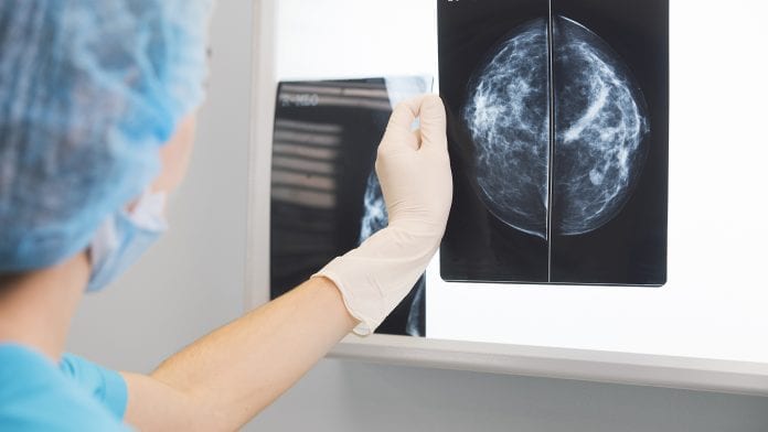
Invasive biopsies may become a thing of the past thanks to a new breakthrough in breast cancer screening.
Harnessing photonics, European scientists have created a new breast cancer screening process using mammographic imaging system that determines benign or malignant breast lesions – spelling an end for unnecessary biopsies and anguish for millions of women.
Scientists from the Horizon2020 project SOLUS have developed a non-invasive, multi-modal, imaging system that uses ultrasound and light technologies to easily differentiate between benign or malignant lesions – without having to perform a biopsy.
Using ultrasound for breast cancer screening
The new screening option is very similar to a pregnancy ultrasound appointment, whereby a clinician scans the breast with a handheld ‘smart optode’ pen probe that combines light and sound to collate blood parameters and tissue constituents.
The scientists use a technique called ‘diffuse optical imaging’ – a method that has provided breakthroughs in neuroscience, wound monitoring, and cancer detection. The scientists can then monitor changes in concentrations of oxygenated and deoxygenated haemoglobin, collagen, lipids and water present in a suspected tumour against a pre-programmed set of results.
Limiting false positive results
While mammography is accurate in detecting breast lesions, many women encounter false positive results – a positive detection of a lump but with no malignant cancer present.
Clinicians, however, do not necessarily know whether a lesion is cancerous or harmless and have to resort to invasive procedures, such as biopsies, to make an accurate diagnosis. According to a recent study, false-positive breast biopsies cost more than €1.85bn ($2bn) per year in the United States alone.
Providing diagnosis
The SOLUS scanner reads a number of different parameters to create a thorough characterisation of tissue, enabling the device to provide a malignant or benign diagnosis
By gathering the total blood volume and oxygenation, collagen, water and lipid content, together with stiffness and morphologic information, the system produces an accurate, while-you-wait diagnosis. Aiming for 95% sensitivity and 90% specificity, the project has combined commercial ultrasound imaging and elastography with novel diffuse optical imaging approaches.
Revolutionising breast cancer diagnosis
The scientists behind the ‘SOLUS’ project are confident their new system will revolutionise breast cancer diagnosis. Professor Paola Taroni from Politecnico di Milano, Italy, said: “Women may have to wait days or weeks for a malignant or benign result to come back, which causes distress, as well as great discomfort from an invasive biopsy.
“Astonishingly, millions of unnecessary biopsies are currently carried out across the world at a cost of millions of Euros in Europe, and potentially billions worldwide.
“Having undergone extensive laboratory trials, the SOLUS team plan to validate the system in real clinical settings at the end of this year and through into 2021.”
Diffuse optics
With ‘diffuse optical imaging’ the system uses light as an investigative tool to look beneath the surface of the skin, without making an incision.
Harnessing photonics and acoustics, this new system gathers diagnostic information from the composition of tissue and blood by sending pulses of infrared light and ultrasound a few centimetres into the breast tissue.
A clinician can then determine whether a lump is benign or malignant given that cancerous tissue is characterised by high haemoglobin and water content, and low lipid content.
“We have been applying diffuse optics to different aspects of breast cancer (lesion discrimination and an estimate of cancer risk) for 20 years now. Our team developed a multi-wavelength time-domain optical mammography which was used in clinical studies on more than 400 patients.
“Now an upgraded version has just entered clinics for the monitoring of neoadjuvant chemotherapy of breast cancer. Furthermore, our group performed a lot of work on other diagnostic applications, such as functional brain imaging and muscle oximetry,” said Professor Taroni.
Peter Gordebeke, Research Manager from the European Institute for Biomedical Imaging Research said: “While SOLUS is developing biomedical imaging technologies to improve the diagnosis of breast cancer, we are making good use of diffuse optics for breast cancer and other medical diagnostics applications.
“SOLUS has created a user-friendly, non-invasive, low-cost, multi-modal imaging system for high-specificity diagnosis of breast cancer.”
“While our multi-modal imaging system can save millions of Euros by reducing the number of unnecessary biopsies, helping women across the world avoid these needless, painful procedures is priceless,” Gordebeke said.
Do you want the latest news and updates from Medical Cannabis Network? Click here for your free subscription, and stay connected with us here.


