Dicronis introduces Lymphit – a cutting-edge technology that will disrupt traditional lymphoedema diagnosis methods for the benefit of cancer survivors.
Patrizia Marschalkova, CEO of Dicronis GmbH (spin-off of ETH Zurich), together with her team, introduces Lymphit, an innovative solution aimed at monitoring the dermal lymphatic function to offer cancer survivors an improved quality of life.
According to the latest and largest global report on cancer (185 countries and 36 types of cancer), published by the International Agency for Research on Cancer (IARC), there were an estimated 18.1 million new cases of cancer and 9.6 million related deaths in 2018. Unfortunately, even for those who defeat cancer, the battle is not over, as there is a long list of post-cancer complications that can further affect their health. Lymphoedema is one of the major long-term complications and a cause of morbidity that is often underestimated. In breast cancer survivors alone, lymphoedema ranges between 14% and 40%.1 The number of patients suffering from lymphoedema and its effects on a person’s quality of life are astonishing. There are over ten million people suffering from cancer-related lymphoedema in the USA and Europe combined.
What is lymphoedema?
There are two types of lymphoedema – primary and secondary. While primary lymphoedema is a genetic condition, secondary lymphoedema usually affects just one limb and occurs after oncologic treatments, whether surgery, radiotherapy or chemotherapy, and for many different types of cancer. It is a chronic, disabling disorder characterised by the progressive accumulation of fluids in tissues. The lymphatic drainage is compromised, and the lymph cannot be pumped back to the bloodstream. The resulting fluid built up in the tissue can lead to more complications, including recurrent skin infections and tissue fibrosis. The most dramatic aspect of the disease is the change in the patient’s physical appearance, which can give birth to devastating psychological traumas.
The main issue with lymphoedema is the lack of an early diagnostic method. In fact, the current methods only manage to diagnose lymphoedema when it’s too late. Proper monitoring of the lymphatic system of cancer survivors is therefore crucial for early diagnosis before the fluid accumulation begins to occur.
Now, medical practitioners rely mainly on symptom analysis and physical examination as a diagnostic method. The main symptoms include transient or persistent swelling, skin changes and recurrent infections. The physical examination consists of assessing the limb size using a tape or its volume by using the water displacement method. In addition to not being highly scalable, both these methods are also imprecise, unreliable, time-consuming and expensive.
Another clinical method consists of an imaging technique called indocyanine green ((ICG) an FDA-approved fluorescent agent) lymphography, which involves injecting a small quantity of ICG solution and using a near-infrared (NIR) camera to visualise the superficial lymphatic structures. The method helps to assess not only whether the patient has lymphoedema but also the stage of its evolution. This method is more accurate but still has significant downsides, including the inability to evaluate the progression of the disease, and a diagnosis when the disease is already advanced. Other limitations of this technique include the need for hospitalisation and a time-consuming and painful procedure.
The most frequent treatment of lymphoedema is called complete decongestive therapy (CDT) and consists of a mix of lymph drainage massage, compression of the affected limb, exercise and self-care. The treatment is not resoluble, but it is most effective when diagnosed in subclinical stages. If diagnosed in a late stage, not only is the efficiency significantly lower but the patient’s quality of life, as well, while the costs of treatment are much higher.
The Lymphit solution
Dicronis plans to solve these issues and offer a solution for all stakeholders involved in the management of the disease. The innovative solution of Dicronis, Lymphit, uses advanced technology to enable a more accurate assessment of the local lymphatic function.
The solution has many benefits for all parties involved. On one hand, patients can benefit from a home-based, pain-free process and a highly scalable diagnosis in the earliest possible stage of secondary lymphoedema. An early diagnosis will further lead to a higher chance for an effective treatment, improved quality of life, and overall patient empowerment. On the other hand, doctors will have better control over the disease and more timely diagnosis.
Physicians will be able to use Lymphit’s insights to adapt the treatment better to the disease evolution and control the therapy outcome more accurately. Lymphit will also determine a better segmentation of patients and lower costs, benefitting health insurances, too.
The technology behind Lymphit is based on the development and use of microneedles loaded with a fluorescent agent to better measure the dermal lymphatic function, aiding both diagnostic and therapeutic processes.
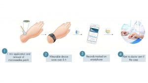
In connection with a wearable device, the solution of Dicronis will further lead to a better quality of life for patients and disruptive innovation in disease management (Fig. 1).

Microneedle patch technology
The human skin is a multi-layered organ with a protective role, guarding the muscles, bones and internal organs from harmful foreign corps. Due to its seven layers, the skin acts as a waterproof barrier, protecting the body from its external environment. The outermost surface layer of the skin (with a thickness of 10-20μm) is called ‘stratum corneum’ and is composed of dead skin cells that block bacteria and prevent them from entering the body.
Microneedles patches are small, micron-sized needles (from 50-900µm) grouped in a large number on a patch that can be applied onto the skin surface, enabling the needles to cross the stratum corneum. This transdermal delivery system is a middle solution between hypodermic injections and transdermal patches. Compared to hypodermic injections, they are much gentler, causing no pain or bleeding, as they don’t affect blood vessels or dermal nerves. Compared to transdermal patches, on the other hand, microneedles manage to ensure contact between a wider variety of substances and the systematic circulation.
Today, microneedles patches are one of modern medicine’s most promising developments, with a cutting-edge technology that is either being evaluated or already in use for drug delivery, vaccination and diagnostic purposes.
There are several types of microneedles being fabricated today. Morphologically, there are mainly four types of needles:
- ‘Poke and patch’– solid, metal needles that allow drugs being applied through a transdermal patch to penetrate the skin
- ‘Coat and poke’ – solid microneedles coated with a drug solution
- Hollow microneedles – these transmit the drug through the cavities they are made of
- Dissolving microneedles – made of biodegradable or water-soluble materials that encapsulate the drug.
Dissolving microneedles
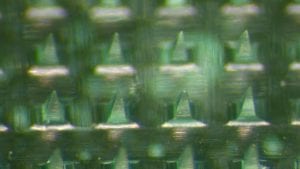
Dicronis chose to develop and use a proprietary dissolving microneedles patch, whose main structure is made of the biocompatible polymer polyvinylpyrrolidone (PVP) and ICG, the NIR fluorescent agent already in use for imaging applications (Fig. 2).

In fact, this choice presents promising features and has multiple benefits. First, the production of dissolving microneedles involves easy manufacturability and low costs but also better control over the release of the substances. These types of microneedle do not leave any sharp materials behind, as the microneedles on the patch dissolve into the skin.
If compared to hollow microneedles, dissolving microneedles do not require qualified personnel, because they can be self-administered, are cost-effective, and offer better stability, as well as the possibility to administer low dosage.
The dissolving microneedle patch of Dicronis allows for simple, painless delivery of the fluorescent agent ICG into the epidermis. Depending on the interstitial location of the ICG-PVP depot, the agent is cleared selectively by the lymphatic system. By measuring the fluorescence at the delivery site over time, the clearance rate of the agent can be evaluated (Fig. 1).
ICG has been used safely for decades in the assessment of hepatic blood flow, cardiac output, ophthalmic angiography and liver function, and is currently the only FDA-approved NIR imaging agent. ICG is also used off-label for image-guided oncology surgery, such as intraoperative identification of solid tumours.2
Microneedles represent the optimal transdermal delivery solution for ICG, but their design is a complex process. Microneedles need to meet several criteria, including water solubility, safety, inertness and mechanical strength. For these considerations, dissolving microneedles are primarily made using polymers and sugars. PVP is one of the polymers that presents several advantages, as its formula not only checks all the previously mentioned criteria but is also crucial for the improved properties of ICG in Lymphit.
Fluorescence and NIR spectroscopy
Before going into more detail about Lymphit, it is worth elucidating the physical properties it makes use of. Light is measured in nanometres and is defined by frequency and wavelength. The different wavelengths of energy produced by a light source create what we call the light spectrum. We can only see the light within a certain range of frequencies and wavelengths, in the part of the spectrum from 380nm to 780nm. The rest of the spectrum comprises of light waves that are not visible to the naked eye, such as X-rays, infrared, UV and NIR.
Fluorescence is the emission of light produced by a fluorescent substance that has absorbed light in a specific range. The NIR window consists of the wavelengths that have the maximum depth of penetration in tissues. NIR fluorescence has several medical applications, as it is minimally absorbed by water and haemoglobin, so the spectra readings can be easily collected from the surface of the skin. NIR spectroscopy is an essential facilitator in both diagnosis and treatment of diseases but also in assessing their pathophysiology. An interesting and innovative approach is the use of NIR light-responsive microneedles for on-demand transdermal drug delivery.
These are some of the principles at the base of Lymphit. The team of Dicronis GmbH aims at translating a technology, selected among the five best inventions of 2015 at ETH Zurich, into a commercially viable product that will drastically improve the overall quality of life of millions of cancer survivors. Lymphit is made solely of PVP and ICG, which are FDA-approved inactive and fluorescent agents respectively.
Lymphit’s focus remains patient protection, so both ingredients are safe and are used in doses that are much smaller compared to the ones used in clinics. This unique combination has multiple advantages, leading to an improvement in their properties, including fluorescence intensity, local kinetics and tensile strength. Overall, this technology will help with functional tissue analysis and serve as an analytical diagnosis tool.
Coupling the microneedle patch to a wearable device
For NIR spectroscopy to enable the subclinical diagnosis, therapy monitoring and pathophysiology assessment in the cases of lymphoedema, the microneedle patch is coupled with a wearable device that measures the fluorescence signal decrease.
The principles behind the wearable device are similar to smartwatches that monitor the heart rate using green LEDs. The green LED lights are paired with light-sensitive photodiodes to illuminate the skin and measure the changes in light absorption. The device therefore manages to detect the amount of blood flowing through the wrist at any moment and hence derive the heart rate.
The wearable device proposed by Dicronis has a similar logic behind it, the only difference being that the light used is at a specific wavelength that makes it compatible with the fluorescent agent. The device will come equipped with LEDs emitting light and photodetectors detecting light in the NIR range. The app and software are currently being designed. Dicronis aims for an app and software that are user-friendly and straightforward, as they will be the user interface for both patients and doctors.
To better understand how this ties to the lymphatic system and its functionality, a better understanding of its role and how it works is needed. The lymphatic system is a network of organs and tissues responsible for fluid haemostasis. The lymph is a fluid that contains white blood cells that fight infections. The fluid is transported through the lymph vessels, which connect to lymph nodes. The nodes help filter the lymph fluid, enabling the body to get rid of toxins and other unwanted materials. The lymphatic system acts as an immune surveillance system, too.
Dysfunctions of the lymphatic system are associated with pathological conditions. A frequent lymphatic dysfunction in cancer survivors is lymphoedema, which manifests itself through swelling of a particular limb. In order to improve disease management and therapeutic interventions, the lymphatic system needs to be understood in correlation to pathological conditions such as lymphoedema but also transplant rejection or cancer metastasis.
Lymphit: microneedle patch, wearable device, software and app
In a medical setting where lymphoedema diagnosis methods are costly, painful and, more often than not, late, Dicronis introduces Lymphit, an innovative product that enables home-based self-administration, a cost-effective and pain-free diagnosis method, and the earliest possible lymphoedema detection system.
The Lymphit system comprises a minimally invasive delivery of the NIR fluorescent agent ICG in the epidermis of the skin through polymeric dissolving microneedle patches. The process is quick (one minute), painless and can be performed at home by the patient. After the patch has been applied, the lymphatic function will be assessed through optical measurements during a period of six hours after the patch application, and the use of a custom wearable device. The latter manages to store data that both the patient and the doctor can access remotely. The evolution can be efficiently monitored on an app the patient can install on their smartphone (Fig. 1).
Lymphit fits perfectly in the digital health domain, owning a unique product that matches the emerging microneedle technology with a smart, wearable device. This digital solution is disrupting the field for lymphoedema diagnosis, offering a long list of benefits, including healthcare cost reductions, patient empowerment, flexibility, early home-based diagnosis, and a personalised treatment and therapy plan.
The research status of the Lymphit project and next milestones
Up until now, Dicronis has raised a total of €550k in mostly non-dilutive financing. In 2016, CEO Patrizia Marschalkova was granted the prestigious ETH Pioneer Fellowship (€130k), with the aim to translate her promising research results into a viable product for commercialisation. Just recently, the company was awarded a grant from Phase I of the H2020 SME Instrument; it is now preparing for Phase II.
So far, the technical focus has been on the development of the microneedle technology. The proof of concept in vivo has shown the system is suitable for assessing the lymphatic function.3 The promising technology is protected by patents filed in Europe, the USA, Japan and Canada. Additional focus has been on regulatory matters and a preliminary market validation through discussions with key opinion leaders; two of them are already on board, and the forecast is very positive.
Moving forward, Dicronis will propose to the regulatory authorities to perform a proof of concept in human subjects to show sustainability in lymphatic function assessment and a larger study to demonstrate the sensibility and specificity of Lymphit.
Before starting the test of the microneedle patches on humans, a regulatory-compliant manufacturing process is the first step. Since the microneedles will enter into the epidermis of the skin, the first requirement is for them to be sterile. Several steps have already been taken in the direction of sterile manufacturing, but when translating the manufacturing from the lab to a small GMP scale, the team is relying on the expertise of industrial partners.
In order to fund the next 18 months of clinical development, securing a €2m investment is crucial. The money is needed to establish the GMP manufacturing and apply the documentation for clinical investigations and the first test on human subjects. Lymphit’s go-to-market strategy will bear significantly lower risks compared to typical healthcare products as it will envisage significantly fewer resources.
The Dicronis team

The Dicronis team (Fig. 3) behind the Lymphit project is led by Marschalkova, who graduated in pharmaceutical sciences and originally developed the microneedle technology during her master’s studies at ETH Zurich. Currently, she is responsible for fundraising, business strategy and project management.
Jovan Jancev, also a graduate of ETH Zurich, is responsible for the R&D Department, where his knowledge and hands-on experience gained from working at an early-stage pharma start-up are put to good use.
Fabrizio Esposito, the current technical director R&D at Gfk, completes the team by bringing his cutting-edge technology expertise, his scientific knowledge, and his business acumen to the table.
Their combined experience and knowledge have led to the creation of a medical technology that will disrupt the market for lymphoedema diagnosis and lymphatic system assessment, for the benefit of cancer survivors.
References
- Rockson (2018). Lymphoedema after breast cancer treatment. N Engl J.doi:10.1056/NEJMcp1803290
- Schaafsma BE, Mieog JS, Hutteman M, van der Vorst JR, Kuppen PJ, Löwik CW, Frangioni JV, van de Velde CJ and Vahrmeijer AL (2011). The clinical use of indocyanine green as a near‐infrared fluorescent contrast agent for image‐guided oncologic surgery. J Surg Oncol 104: 323-332. doi:10.1002/jso.21943
- Brambilla D, Proulx ST, Marschalkova P, Detmar M and Leroux J (2016). Microneedles for the Noninvasive Structural and Functional Assessment of Dermal Lymphatic Vessels. Small 12: 1053-1061. doi:10.1002/smll.201503093
Patrizia Marschalkova
Dicronis GmbH
+41 44 633 63 19
patrizia@dicronis.com
www.dicronis.com
Please note, this article will appear in issue 8 of Health Europa Quarterly, which is available to read now.


