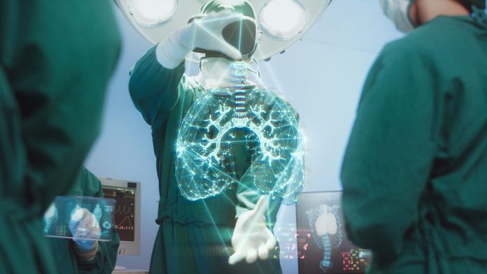
Scientists at King Abdullah University of Science and Technology (KAUST) have developed an Artificial Intelligence (AI) tool that can accurately diagnose COVID-19 lung damage.
The cutting-edge technology is a significant advancement on traditional diagnostics, which are both time-consuming and unreliable. The novel KAUST method is called Deep-Lung Parenchyma-Enhancing (DLPE). The tool effectively identifies COVID-19 lung damage by overlaying AI algorithms on top of conventional imaging data, revealing visual features of lung dysfunction that would otherwise be undetectable.
Xin Gao, a computer scientist and computational biologist at KAUST, commented: “Through DLPE augmentation, radiologists can discover and analyse novel sub-visual lung lesions. Analysis of these lesions could then help explain patients’ respiratory symptoms, allowing for better disease management and treatment.”
Limitations in identifying COVID-19 lung damage
COVID-19, similarly to other respiratory infections, can cause long-term lung damage; however, it is currently difficult to visualise this damage. This is due to conventional chest scans not being able to detect signs of lung scarring and pulmonary abnormalities reliably. Subsequently, this makes monitoring the health and recovery of COVID-19 patients with long-term breathing difficulties and other post-COVID complications challenging.
AI diagnostics for COVID-19
The team’s new AI tool works by initially eliminating any anatomical features that are not associated with lung parenchyma – the tissues that are instrumental in gas exchange and act as the primary sites of COVID-19 lung damage. During this process, the airways and blood vessels are removed from the images, with the pictures then enhanced to expose lesions that would have persisted unidentified without the AI technology.
The KAUST scientists trained and refined their AI algorithms by employing computed tomography (CT) chest scans from thousands of patients in China who were hospitalised with COVID-19. The team then optimised the tool with input from expert radiologists and applied DLPE to various survivors of COVID-19 who were still experiencing lung problems. All of the patients had suffered severe SARS-CoV-2 infection and required intensive care treatment.
This analysis illuminated that DLPE can effectively highlight signs of COVID-19 lung damage, such as pulmonary fibrosis, in patients with long-term complications, helping to explain their shortness of breath, coughing, and other respiratory issues. Gao expressed that this diagnosis would have been impossible using standard CT image analytics.
He said: “With DLPE, for the first time, we proved that long-term CT lesions could explain such symptoms. Thus, treatments for fibrosis may be very effective at addressing the long-term respiratory complications of COVID-19.”
Detecting other lung problems
Although DLPE was initially designed to monitor recovery following COVID-19 infection, the team also utilised the tool on chest scans obtained from patients with a variety of lung problems, such as pneumonia, tuberculosis, and lung cancer. This additional testing demonstrated that DLPE could be used as a broad diagnostic tool for all lung conditions.


