A group effort between physicists, biologists and engineers has led to a novel method to analyse a key protein in hepatitis C virus. Collaborators hope this will lead to better targeting mechanisms for future drug development.
The blood-borne hepatitis C virus (HCV) is having a considerable impact on people’s lives worldwide. As the virus is a cause of liver disease and cancer, sadly more than 300,000 people die each year after contracting it. This is while 71 million people continue to live with a chronic infection, at risk of it developing into something more serious.1 While antiviral medicines are currently used to treat hepatitis C, research groups are still on the hunt to discover more about the inner workings of the virus. Understanding how the virus spreads can help to identify opportunities to prevent it from doing so, and potential targets for new therapies.
One such candidate is the p7 protein. It plays a key role in the release of the Hepatitis C virus, influencing its lifespan and persistence in the human body, as well as how the disease continues to develop while it is there – as such, it is an ideal target to consider for the development of novel treatments for people who have contracted the virus.
However, little data is currently available on the p7 protein, and, up until now, various factors have hindered our ability to analyse how it interacts within its environment. No less because it is extremely difficult to get hold of a sufficient amount for experiments – expression of the p7 membrane protein in classical overexpression systems is difficult due to its high content in hydrophobic amino acids and its small size. Both functional and structural studies require a substantial amount of the protein, which is extremely challenging due to the small quantities produced. Current methods for protein production require tremendous work and display some limiting features on numerous aspects.
Old challenges lead to a new revelation
As well as limited access to the protein itself, structural analysis of p7 has been further restricted because it is difficult to crystallise. There are challenges at all levels of the process, including expression, solubilisation, purification, crystallisation, data collection and structure solution.
In the first instance, to produce crystals requires that the proteins have been stable for hours, and sometimes even months. However, unfortunately, it is not often the case that membrane proteins (p7 is such a protein) are stable enough to do this. This is because a crystal structure depends mostly on hydrophilic intermolecular interactions (between proteins) while membrane proteins like p7 have mostly hydrophobic surfaces. The detergents used to stabilise membrane proteins often interfere with the crystallisation process, making it hard to get and reproduce. Finally, having produced a crystal does not necessarily mean that it is one fit for purpose. Crystallisation campaigns are costly and time consuming, with no results guaranteed.
For this reason, the field has generally looked towards alternative methods to analyse the structure of the protein, including nuclear magnetic resonance (NMR) techniques. While this method is achievable, up until now analysing the p7 protein this way has remained insufficient to guide novel therapies. This is because it is a membrane protein, and so sits in a specific lipid environment, which also needs to be considered. Solid state NMR studies are limited in this respect, making it difficult to observe the protein in enough context to guide research.
That is until a collaboration between the Institut Laue-Langevin (ILL), Synthelis SAS and University Grenoble Alpes, France, set out to develop a novel method to study the protein’s structure integration within its native membrane environment using neutron reflectometry (NR).
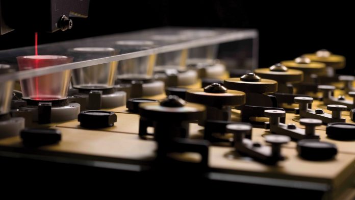
We opted to use a time-of-flight reflectometer, named FIGARO, at the ILL in Grenoble, the world centre for neutron science, to perform the experiment – ideal, as it is optimised for the study of soft matter and biological molecules at interfaces. We formed bio-membranes supported on quartz crystals and used FIGARO to transmit an incident neutron beam through the substrate, and reflect from the solid-liquid interface.
ILL’s FIGARO is able to strike the interface from above or below the sample in a wide q-range of up to 0.42 Å-1 (reflection up) or 0.27 Å-1 (reflection down) for horizontal samples, although we did not need such great ranges. Analysing the p7 protein using FIGARO revealed momentum transfer ranges of 0.008> qz> 0.2 Å-1 and minimum reflectivities of R ~ 5×10⁻⁷ using wavelengths λ = 2-20 Å, as well as two angles of incidence, and a dqz/qz resolution of 10%.
We were excited to observe that this process, for the first time, revealed the structure of a functional p7 protein complex from HCV within a physiologically relevant lipid bilayer, at nanoscale resolution.
Turning structure into treatments
As published in a Nature Scientific Reports paper, this technique, combined with cell-free protein expression performed by Synthelis, was able to show that the p7 protein from HCV assembles within the lipid membrane into oligomers that take the shape of a funnel.2 Observing this conical shape told us the p7 protein preferred protein orientation, as well as its specific insertion mechanism – helping to outline the precise target mechanisms for future drug development.
Complementing what researchers had already been able to decipher about the importance of the protein for HCV, the study has further paved the way for the in silico development of new drugs that can target p7 and thus the virus, and is therefore on the way to helping establish treatments that are more effective. A new class of treatments would also be most useful for those patients that are yet to respond to the current options.
Beyond HCV
In addition to this, a deeper understanding of the virus will not only help identify treatments but can also help to support the continued development of novel nanostructured systems and devices. This technology lies at the heart of creating efficient biosensors, which can be used for in vitro diagnostic and biotechnological applications. Helping in turn to better diagnose patients and identify appropriate opportunities for treatment.
This discovery is not limited to HCV, as the method itself can translate to other areas of discovery. For instance, membrane protein dysfunction is also correlated with a wide range of diseases. This involves the defective transport of small molecules and ions within the membrane, so the advancement in methods and ability to analyse membrane proteins in their native condition, at an atomic scale, has the potential to help support new therapeutic approaches in these other areas, too, including the development of antibodies against HIV.
Beyond this, the behaviour of membrane proteins in lipid bilayers can also be useful in other fields, beyond the life sciences sector. It can be used as a tool to understand membrane systems used in biofuel cells, or even the technology using membrane proteins for water filtration.
As illustrated by this work, the ongoing cross-sector collaboration between physicists, biologists and engineers from these organisations in Grenoble has proven an important opportunity to take the fundamental understanding of physical and biological processes that underlie the development of various nanostructured systems and devices, across many fields and industries.
References
- http://www.who.int/mediacentre/factsheets/fs164/en/
- Coupling neutron reflectivity with cell-free protein synthesis to probe membrane protein structure in supported bilayers, Thomas Soranzo, Donald K Martin, Jean-Luc Lenormand, & Erik B Watkins [doi: 10.1038/s41598-017-03472-8]
Erik Watkins
Former ILL Instrument Scientist
Thomas Soranzo, Donald Martin and Jean-Luc Lernomand
University Grenoble Alpes
Bruno Tillier
Managing Director
Synthelis SAS
www.ill.eu
www.univ-grenoble-alpes.fr
www.synthelis.com
This article will appear in issue 4 of Health Europa Quarterly, which will be published in February.


