Researchers from Linköping University have found that there are major biological differences between dense breast tissue and non-dense breast tissue, suggesting that dense breasts promote cancer growth.
Currently, dense and nonsense breasts are treated identically in the Swedish healthcare system. However, recent findings have shown that the risk of developing breast cancer could be up to five times higher than those with dense breast tissue.
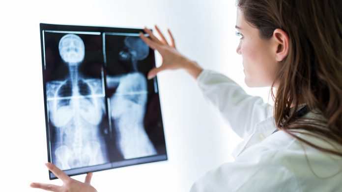
All women over the age of 40 in Sweden are regularly offered mammograms, which uses magnetic resonance imaging (MRI) and ultrasound to detect tumours in the breast. Dense breast tissue appears white in mammograms, whereas non-dense breast tissue appears grey.
“The problem is that we don’t know what to do with the women who have dense breasts. Large studies would be needed before introducing a screening programme for such women, such that we can identify those with the greatest risk and follow them in the healthcare system. This is necessary to prevent putting many women through unnecessary examinations,” explained Charlotta Dabrosin, professor in the Department of Biomedical and Clinical Sciences at Linköping University.
Tumours are harder to find in dense breast tissue
Breast density relates to the making of an individual’s connective tissue. Both glandular tissue and connective tissue appear white in mammograms, meaning they can be more difficult to detect by mammography. However, the difficulty in tumour detection does not fully explain the higher risk of cancer in women with dense breast tissue. The other risk factors are currently unknown.
The researchers decided to investigate to what extent the biological properties of dense and non-dense breasts differed. They did this by developing an MRI method that could measure breast density and other distinguishing factors of breasts more accurately than existing methods.
The researchers examined 44 women with varying breast densities using contrast-enhanced MRI. The research team also used microdialysis, a technique where a thin catheter is inserted into the breast tissue to obtain samples of the fluid that surrounds the cells. Previous studies have shown that the microdialysis in dense breasts is very similar to that in breast tumours.
The study found unexpectedly large differences between healthy dense and non-dense breasts. The researchers measured the levels of 270 proteins and found that the levels of 124 of them were elevated in dense breasts. These proteins have been linked with cancer development through underlying processes such as inflammation, the formation of new blood vessels, and cell growth.
Understanding biological differences in tissue can lead to better treatment
“There are huge biological differences between dense and non-dense breasts. What I find amazing about our results is that we can link the levels of proteins such as inflammatory proteins and growth factors with the differences in breast physiology that we showed using MRI. We found, for example, that the contrast agent diffuses differently in the different types of breasts, which suggests that the blood vessels are affected,” said Dabrosin.
While the researchers have observed clear correlations between breast density and cancer risk, they have not identified the cause. However, the links between the amounts of protein and physiological differences are strong enough for the researchers to conclude that the links are causal.
Approximately one in three women aged 40-50 has precursors to cancer in their breasts. Fewer than 1% of women in this age group develop cancer as the growth remains at this early stage. The researchers believe that dense breasts may have an advantageous microenvironment that promotes the transition of anomalous cells into cancer.
“The results raise many questions about whether it is possible to reduce the levels of these proteins and reduce the risk of developing cancer. We open possibilities that we have not previously had,” concluded Charlotta Dabrosin.


