A new study has revealed insights into the stem cell microenvironment through 3D imaging technology.
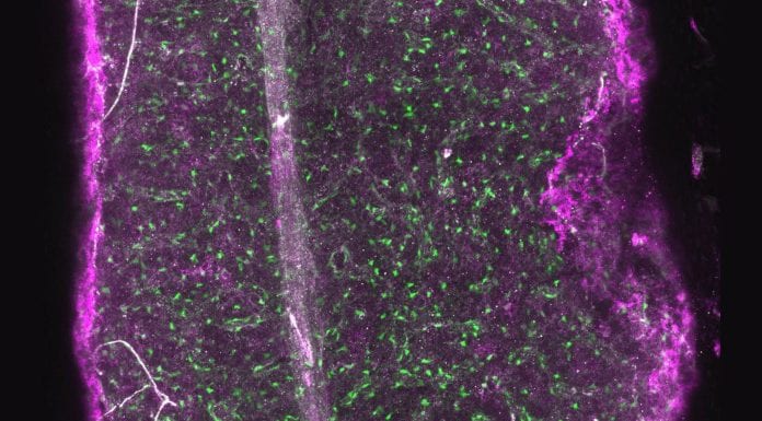
Researchers from the European Molecular Biology Laboratory (EMBL) and the German Cancer Research Center (DKFZ) in Heidelberg, Germany, have developed new methods to reveal the three-dimensional organisation of bone marrow at a single cell level.
Since bone marrow harbours blood stem cells responsible for life-long blood generation, these results and the new method provides a novel scientific basis to study blood cancer.
The results have been published in Nature Cell Biology.
Looking at bone marrow
In the published study the group, led by researchers from EMBL and the DKFZ, present their new methods that permit them to characterise complex organs. The team focussed their research on the bone marrow of mice, as it harbours blood stem cells that are responsible for life-long blood production.
Because of the ability to influence stem cells and to sustain blood production, there is a growing interest in exploiting the bone marrow environment as a target for novel leukaemia treatments.
Chiara Baccin, a post-doc in the Steinmetz Group at EMBL Heidelberg, said: “So far, very little was known about how different cells are spatially organised within the bone marrow and how they interact with each other.”
The new methods permitted the team to uncover the cellular composition, the three-dimensional organisation and how the cells interact with each other. In the process the researchers have identified previously unknown cell types, so called ‘niche cells’, that are of great importance for the regulation of blood stem cells.
In order to understand which cells can be found in the bone marrow, where they are localised and how they might impact on stem cells, the researchers combined single-cell and spatial transcriptomics with novel computational methods.
By analysing the RNA content of individual bone marrow cells, the team identified 32 different cell types, including extremely rare and previously unknown cell types.
Simon Haas, group leader at the DKFZ and HI-STEM, said: “We believe that these rare ‘niche cells’ establish unique environments in the bone marrow that are required for stem cell function and production of new blood and immune cells.”
Novel computational methods
Using novel computational methods, the researchers were not only able to determine the organisation of the different cell types in the bone marrow in 3D, but could also predict how cells interact with each other.
Lars Velten, staff scientist in the Steinmetz Group, said: “It is the first evidence that spatial interactions in a tissue can be deduced computationally on the basis of genomic data.”
The data, which is now already used by different teams all over the world, is accessible in a 3D-atlas via a user-friendly web app. All gathered information is available for free and will in the future facilitate the refinement of genetic studies.
The developed method can be used in different tissues for the study of their 3D organisation.
“Our approach is widely applicable and can be used to study the complex pathology of human diseases. We are actually currently in the process of using these approaches on haematological diseases, such as leukaemia,” highlights Andreas Trumpp, managing director of HI-STEM and division head at DKFZ.


