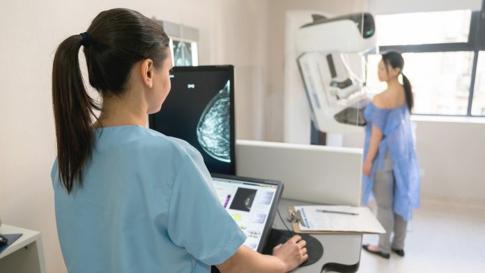
A Norwegian study suggests that Artificial Intelligence (AI) may be effectively used to perform breast cancer screening, potentially speeding up diagnostics for the disease.
The new research is the most extensive to date and was pioneered by a team from the Cancer Registry of Norway. For their study, the investigators evaluated the performance of a commercially available AI system against a routine independent double reading performed in a traditional population-based breast cancer screening programme.
The findings of the study are published in Radiology.
AI breast cancer screening
Mammograms obtained in population-based breast cancer screening programmes provide a substantial workload for radiologists, meaning novel methods for making this process more efficient could save time and save lives due to a rapid diagnosis. AI has been suggested as an automated second reader of mammograms to alleviate this workload and has shown great potential for cancer detection; however, there is limited evidence for its performance in actual screening settings.
To test the proficiency of AI, the team utilised data from around 123,000 examinations conducted on more than 47,000 women at four facilities in BreastScreen Norway, the country’s population-based breast cancer screening programme.
The dataset included 752 cancers that were detected at screening and 205 interval cancers (cancers detected between screening rounds). The AI technology predicted cancer risk on a scale from one to ten, with one being the lowest risk and ten being the highest. The team identified that 653 of the 752 (87.6%) screen-detected and 92 of 205 (44.9% of interval cancers had the highest AI score of ten.
Next, the team established three thresholds to evaluate the performance of the AI system as a decision-making tool. The researchers developed a threshold that mirrored the average rate at which a radiologist makes a positive interpretation, finding that the proportion of screen-detected cancers not identified by the AI was less than 20%. Despite the promising performance of AI, more research is needed due to the study utilising retrospective data.
Solveig Hofvind, the leader of the study from the Cancer Registry of Norway, commented: “In our study, we assumed that all cancer cases selected by the AI system were detected. This might not be true in a real screening setting. However, given that assumption, AI will probably be of great value in the interpretation of screening mammograms in the future.”
Advancing diagnosis
The study results demonstrated strong histopathologic characteristics associated with a better prognosis for screening-detected cancers with low versus high AI scores. However, the opposite was found for interval cancers, suggesting that interval cancers with a low AI score are true interval cancers that are not visible on the screening mammograms.
The high percentage of true negative examinations classified with a low AI score may potentially reduce the interpretive volume whilst allowing only a small number of cancers to go undetected. The team explained that by using AI as one of the readers in a double reading setting, radiologists would still be able to identify these cancers.
“Based on our results, we expect AI to be of great value in the interpretation of screening mammograms in the future,” Dr Hofvind said. “We expect the greatest potential to be in reducing the reading volume by selecting negative examinations. We are looking forward to testing out different scenarios for AI using retrospective data and then running a prospective trial.”


