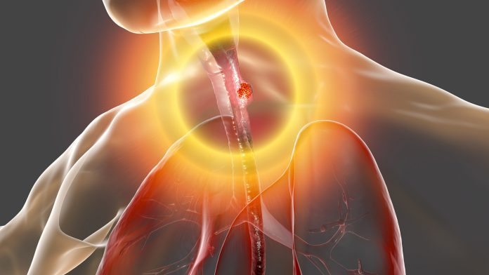
Scientists have created a novel Artificial Intelligence (AI) diagnostic tool that may advance the detection of oesophageal cancer for people with a condition known as Barrett’s oesophagus.
Developed by researchers at the University of Colorado (CU) Cancer Center, the new AI diagnostic platform may revolutionise how healthcare professionals detect oesophageal cancer in patients with Barrett’s oesophagus, significantly improving patient outcomes for the condition.
What is Barrett’s oesophagus?
Barrett’s oesophagus is a premalignant condition for oesophageal cancer and occurs when the flatlining of the oesophagus is damaged due to acid reflux. Barrett’s oesophagus is found in around 7% to 10% of people with chronic gastroesophageal reflux disease (GERD) and is present in 1% to 2% of the general adult population.
Due to Barrett’s oesophagus being linked to an increased risk of developing oesophageal cancer, individuals with the condition should undergo regular biopsies to check for precancerous cells – known as dysplasia – or early-stage oesophageal cancer. If detected early, oesophageal cancer can often be treated endoscopically, whereas it usually requires surgery in later stages with or without chemotherapy or radiation therapy.
Wani said: “Oesophageal cancer is a highly lethal cancer with a five-year survival rate of less than 20%. What is really disappointing is that despite all the advances that we’ve made, the vast majority of patients with oesophageal cancer still present with advanced-stage disease.”
Utilising AI
With sponsorship from medical technology company CDx Diagnostics Inc., Wani’s team will be performing a four-year study of CDx’s advanced diagnostic platform called Wide-Area Transepithelial Sampling with computer-assisted three-dimensional analysis (WATS3D). The researchers will compare the diagnostic test against the current standard of care known as the Seattle biopsy protocol. Whereas the Seattle biopsy protocol includes using four-quadrant biopsies of the oesophagus, the CDx method uses a unique brush instrument that locates more at-risk mucosa.
The team will review specimens at the company’s CLIA-certified laboratory using its patented extended depth of field imaging system and AI platform that enables diagnostic precision. The investigation will be conducted at 14 centres across the US, with the primary aim being to examine the diagnostic yield of dysplasia or cancer of the two methods.
Wani commented: “It’s a computer-assisted, three-dimensional analysis of samples that we obtain during endoscopy from the Barrett’s segment. Instead of using forceps biopsies, it’s a brush device that allows you to extensively sample the Barrett’s segment. Then, using the synthesised 3D images and neural network analyses, these samples get assessed, and abnormal cells get flagged for the pathologist to look at. The goal is to find patients early in their progression so that they can avoid having to go through chemotherapy, radiation, and esophagectomy, which are treatments we reserve for advanced-stage cancers.”
Optimising cancer detection
The team explained that it is essential to investigate Barrett’s oesophagus due to being the only identifiable premalignant condition for oesophageal cancer. Moreover, traditional biopsies may miss as much as 30% of patients with Barrett’s oesophagus-related dysplasia or early-stage oesophageal cancer. They hope their study will improve diagnostics to identify the disease before it progresses.
“We are excited to be embarking on such an important study that really will impact thousands of patients that are diagnosed with Barrett’s oesophagus and oesophageal cancer,” Wani said. “If the study shows that this sampling method detects more patients with dysplasia and early oesophageal cancer and improves outcomes, this could have significant ramifications for the way we perform endoscopy in the future for this patient population.”


